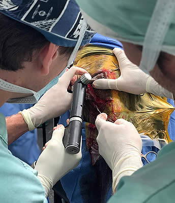
Dr. Michael Myrhe and Dr. Weston Davis performing the Ethmoid Hematoma procedure. Photo courtesy of PBEC.
A horse was recently admitted to Palm Beach Equine Clinic (PBEC), based in Wellington, FL, with symptoms that included bleeding from the nostril. The patient’s referring veterinarian had diagnosed the horse with an ethmoid hematoma, which in layman’s terms is essentially a mass that fills with blood in the nose or sinus cavity.
The patient was placed under the care of PBEC’s board-certified surgeon Dr. Weston Davis and Dr. Michael Myhre. They performed an airway endoscopy to locate and evaluate the hematoma that the referring veterinarian had identified. After confirming the diagnosis, Dr. Davis and Dr. Myhre were eager to ensure that it was the one and only hematoma they were battling.
PBEC is one of an elite group of equine veterinary clinics to have a computed tomography (CT) machine in their arsenal of diagnostic imaging equipment. A CT gives veterinarians a unique look at the head, neck, and spine of a horse that they would never be able to accomplish with other imaging modalities. After a CT of the patient’s sinuses, more masses were indeed identified.
“This was a fairly typical presentation of an ethmoid hematoma, but there were certainly more masses than normal,” said Dr. Myhre. “It’s for this reason that the CT was very useful. If we were not able to obtain the scans that we did, we may have missed the masses that were located deeper in the sinus.”
Click here to watch the CT scan that spotted the additional masses in progress.
The cause of an ethmoid hematoma is unknown, but the mass resembles a tumor in appearance and development without being neoplastic. Horses with extensive masses may have reduced airflow and an expanding hematoma can cause pressure necrosis of the surrounding bones, but rarely causes facial distortion. Treatments of the condition can range from conservative management to surgery. The conservative treatment route includes the injection of formalin – a mixture of formaldehyde gas and water – into the mass using a guarded endoscopic needle. Once injected, the mass typically regresses rapidly, but recurrence is common. For some cases, surgical excision is achieved via a frontonasal bone flap procedure.
Due to the location and advances nature of the masses in this case, injection was not an option and the CT imaging was used to plan a surgical approach. “After sedation and a local block, we went into the sinus through a flap approach where we took a section of bone, cut it into a flap, and moved it back so we could go into the sinuses through a nice window,” said Dr. Myhre. “We removed a mass four centimeters in diameter as well as several smaller masses two to three centimeters in diameter, then flushed the area and closed.”
According to Dr. Myhre the advantages of a standing procedure included fewer risks from bleeding and fewer risks of recovering from anesthesia.
Post-surgery, the bone flap will require several weeks to heal, but the skin itself healed within one to two weeks, which is when the horse was cleared to return to normal activity.
Jennifer Wood, Jump Media
jennifer@jumpmediallc.com